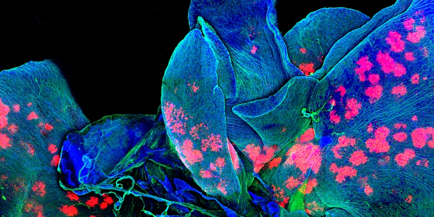

The Microscopy Core Facility provides experimental consultation, fee-for-service imaging assistance, and training in and access to several imaging platforms, image acquisition equipment, and data analysis software packages.
Services include:
- consultation on fluorophores and experimental design
- light and epifluorescence microscopy
- fluorescent slide scanning
- scanning confocal microscopy
- multiphoton scanning confocal microscopy and intravital imaging
- deconvolution-based microscopy for sensitive, high-resolution fluorescence imaging
- 3D structured illumination microscopy
- total internal reflection fluorescence (TIRF) microscopy
- other advanced fluorescence microscopy techniques such as fluorescence recovery after bleaching (FRAP) and Förster resonance energy transfer (FRET)
Access is typically reserved for KI members. In some circumstances, access may be available to non-member MIT or external users (details available on request from the Scientific Director, Jeffrey Kuhn).
The Microscopy Core is supported in part by funding provided to the Koch Institute from a National Cancer Institute Cancer Center Support Grant.
