

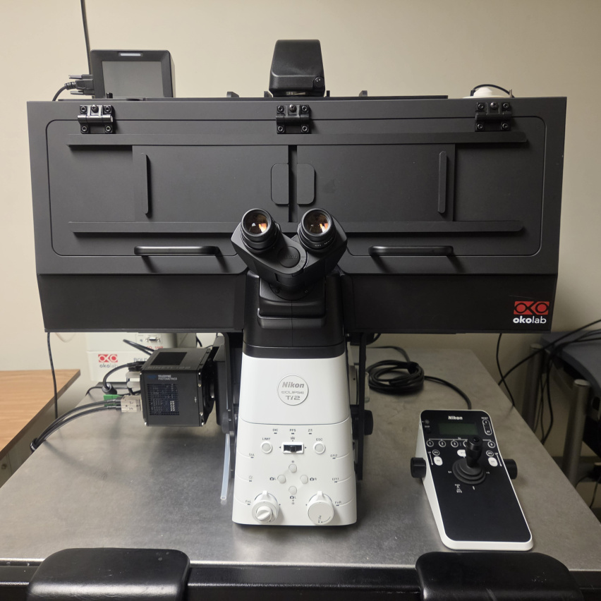
Overview
- Fast Fluorescence Microscope for long-term 3D live-cell acquisition
- BL1 samples ONLY (fixed samples or live non-human material or cell lines)
- BL2 imaging coming fall 2026 (live human-derived cells and tissues)
Location
- Located in the Building 68 Bio-Core Satellite Facility
- Contact KIMCF-68 staff Linda Melodia for assistance
Specifications
- Inverted microscope with automated XY stage and Z axis
- Well-plate scanning
- Single chamber and 4 slide adapters available
- Heated environmental chamber with CO2 for live cell imaging
- Perfect Focus system for long-term scanning
- Lumencor CELESTA laser epifluorescent-illumination
- Critical "tophat" illuminator for fast, even excitation
- LED transmitted light source
- Motorized condenser turret
- 7 fluorescent channels with fast emission filter wheel
- Sensitive back-thinned Teledyne Kinetix 22 sCMOS camera
- Nikon Element software with General Analyis
- Jobs visual programming for complex acquisitions
Fluorescent Filters
| Name | Set | Std dyes | Cy series | Alexa | Ex | Em |
|---|---|---|---|---|---|---|
| DAPI | Penta | DAPI/Hoechst/BV421 | 405 | 432/36 | ||
| FITC | Penta | FITC/GFP | Cy2 | AF488 | 488 | 525/40 |
| TxRed | Penta | TRITC/TxRed | Cy3.5 | AF546/594 | 561 | 595/31 |
| Cy5 | Penta | APC | Cy5/Cy5.5 | AF647 | 640 | 680/42 |
| Cy7 | Penta | Cy7 | Cy7 | AF750 | 730 | 809/81 |
| CFP | CFP/YFP | CFP | 440 | 485/20 | ||
| YFP | CFP/YFP | YFP/TRITC | AF514 | 514 | 560/25 |
Objectives
| Objective | OIL | Trans | Corr | WD mm | Z DOF um |
|---|---|---|---|---|---|
| 4x/0.13 | PhL | Plan Fluor | 17.1 | 45 | |
| 20x/0.8 | DIC | Plan Apochromat Lambda D | 0.8 | 1.27 | |
| 40x/0.75 | Ph2 | Plan Fluor | 0.66 | 1.19 | |
| 60x/1.42 | Oil | DIC | CFI Plan Apochromat Lambda D | 0.13 | 0.53 |
Data Transfer
- USB flash storage or drives are NOT PERMITTED on core instruments.
- All data must be transferred over the network
- Use the nearby workstation in office 68-371 to transfer your data from the microscope to your server