
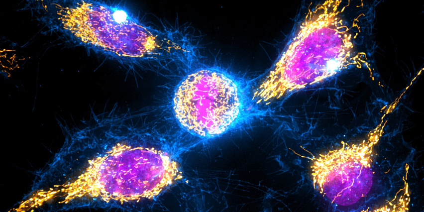
Welcome to High Throughput Sciences: High Content Imaging, a portfolio of shared resources within the Koch Institute High Throughput Sciences Core that provides Koch Institute and MIT researchers access to a range of longitudinal live cell imaging and analysis technologies.
The Core offers access to the following tools:
- Incucyte S3 system that allows automatic image acquisition and analysis of cells in a physiologically relevant environment.
- Nanolive 3D Cell Explorer 96focus allows automated, label-free live cell imaging and analysis under ambient conditions.
- Revvity Operetta CLS High-Content Microscope, an environmentally-controlled high-content microscope integrated with the HTS automation platform.
- CellCelector, a tool which enables users to isolate single cells using an ultra-precise robotic micromanipulator. We also offer access to nanowell microplates which enable automated isolation of monoclonal and viable clones.
- Genepix 4400 Laser Scanner can be used to scan slide based microarrays. Available lasers: Red, Blue, Green & Yellow (635, 488, 635a and 594nm).
Access to the High Throughput Sciences High Content Imaging resources is available to all members of the MIT community, to the extent permitted by available capacity. Priority access is given to KI members, NCI-funded research projects and other contributing user groups in recognition of funding support. Depending on available capacity, access may be available to non-MIT users (details available on request from Live Cell Imaging Scientist Shanu Mehta).
The High Throughput Sciences Core is supported in part by funding provided to the Koch Institute from a National Cancer Institute Cancer Center Support Grant.